Viruses
Page 6
![]()
 |
Section Objectives · Describe the structure of a bacteriophage. · Name the five phases of the lytic cycle. · Explain how the process of transduction occurs. · State a possible theory for the evolution of viruses.
|
Reproduction
and Evolution
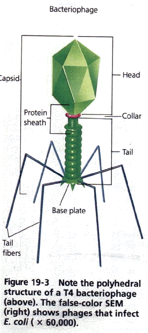
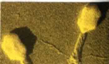
Scientists first learned about virus reproduction by studying bacteriophages, viruses that infect bacteria. Phages (PA Yiuz), as they are called, can be easily studied because their bacterial hosts multiply quickly in cell cultures. The most commonly studied phages are those of the T group. They are named Ti, T2, T3, and so forth. The Tphages infect the bacterium Escherichia coli, the common bacterium of the human digestive tract. The T-even phages (T2, T4, T6) are virulent. They are capable of destroying E. coli cells. Examine the structure of the T4 phage in Figure 19-3. Notice that its morphology is different from that of the viruses described in Section 19.1. DNA in the viral core is surrounded by a protein coat that forms a polyhedron. Beneath the head is a collar of protein and a sheath that rests on a base plate. Tail fibers emerge from the base plate. As you read about the reproduction of the T4 phage, notice how the structure of the virus suits its function. The Lytic Cycle The lytic cycle is a fundamental reproductive process in viruses. The term lyse means to "break open," a reference to the liberation of the new viral particles from the host cell. The T4 phage reproduces by the lytic cycle and thus can serve as an example of viral reproduction. The lytic cycle has five phases, each of which is continuous with the others. The phases are adsorption, entry, replication, assembly, and release. Study Figure 19-4 as you read. 1. During adsorption the virus attaches itself to a specific host cell. The tail fibers of the virus contain proteins that have a chemical affinity with the bacterial cell wall. In fact, specific areas of the wall, called receptor sites, are the places where the virus attaches itself. 2. During entry the T4 phage releases an enzyme that weakens a spot in the cell wall of the host. Then, much like a hypodermic needle, the T4 presses its sheath against the cell and injects its
|
 |