Viruses Page 3
![]()
 |
 |
infecting
a tissue culture of living cells with a strain of
a virus, researchers can grow viruses and examine
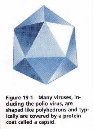
them. Researchers grow large quantities of a virus in tissue cultures and then subject the viruses to serological and electrophoretic studies. Serology (sub-RAHL-uhjee) is the study of biological fluids. In particular, serologists often determine an organism's antibody responses to viruses. Electrophoresis (ih-LECK-truh-fuhREE-suss) is a process that separates molecules, especially proteins, on the basis of their specific electrical charges. Electrophoresis is used to separate and examine the protein components of viruses. In addition, recent advances in DNA and RNA sequencing have enabled virologists to determine the sequence of bases in the viral nucleic acids. Using all these techniques and others, virologists have determined the structure of many viruses. StructureThe structure of the polio virus shown in Figure 19-1 is typical of many viruses. The virus particle is about 20 to 30 nm in diameter. The capsid is shaped like an icosahedron (ie-KOE-suh-HEEdrun)-a polyhedron with 20 triangular faces. The capsid is made of protein subunits that fit together like the pieces of leather on a soccer ball. The capsid surrounds a single strand of RNA. Most icosahedral viruses are between 15 and 200 nm in diameter. The approximately 200 kinds of viruses that cause the common cold are mostly icosahedral viruses about the size and shape of the polio virus. Some viruses, such as the virus that causes tobacco mosaic disease, a disease of tobacco plants, are rod shaped when viewed under the electron microscope. These viruses have a helical strand of nucleic acid that runs the length of the |
 |
|
 |
|
